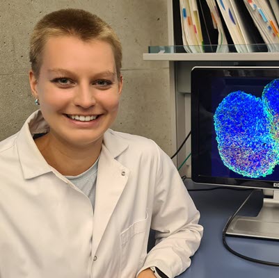
Three years into a medical degree in the UK, Annika Schulz came to UBC to complete an MSc in Microbiology and Immunology in the Jean Lab. Drawn to this path by an interest in virology, she entered the Bioimaging North America Imaging Contest after being initiated to confocal microscopy. A fast learner with a flair for telling detail, Schulz took third place in a highly competitive international competition. The affirmation is a welcome one for Schulz, who states that “capturing intricate details of biological processes in a predominantly non-invasive manner will certainly continue to be central to my work as a future clinician and scientist.”
A Q&A with Annika Schulz
Q: Tell us about the competition! Was this by invitation or submission? Who were you competing against? As a Master’s student, were you up against PIs and PhDs?
A: The BINA (BioImaging North America) Image Contest 2022 was open to all levels of bioimaging scientists in Canada, USA and Mexico. To participate, you had to become a member of the inclusive and very supportive BINA community – it’s free to join! The BINA organization seeks to connect bioimaging scientists and facilities a variety of community events. I did not attend the community conference this year, but I look forward to joining some of their events in future. Currently, I use both the microscopes at the UBC Bioimaging Facility and the LSI Bioimaging Core. The UBC Bioimaging Facility sent out a notice about the BINA Image Contest in September, which is how I came across the competition and decided to submit some of my images.
To date, I have completed the first half of my Bachelor of Medicine, Bachelor of Surgery degree (MBBS) at University College London, where I will resume clinical training upon finishing my MSc in Microbiology & Immunology at UBC. As an MSc student in the Jean Lab (Department of Microbiology & Immunology, Life Sciences Institute), I work with complex in vitro culture systems including air-liquid interface cultures (human airway model) and induced pluripotent stem cell-derived cerebral organoids (lab-grown mini brains). These cell-based assays allow for physiologically relevant screening of host-directed antivirals and the interrogation of their mechanism of action.
Q: Was the competition virtual or in person? In your image, what did you show, what microscope did you use, and who were your collaborators on the project?
A: The BINA Image Contest was virtual, but the results were announced at the recent community conference. My image entitled ‘Brainstorm’ shows a lab-grown ‘mini brain’. I used fructose-glycerol clearing reagent to achieve tissue clearing after performing immunofluorescence staining. The image was then acquired using the Leica SP5 Inverted Confocal Microscope in the LSI (63X silicon oil immersion) with a tile scan protocol.
Brainstorm – Annika Lea Schulz
Life Sciences Institute Imaging – Advanced Bioimaging Core at University of British Columbia
Sample: Human induced pluripotent stem cell-derived dorsal forebrain organoid with adenoviral-vector mediated expression of neuro-serine protease inhibitor (neuroserpin) Spn4A. Clearing was performed after immunofluorescence staining using fructose-glycerol clearing reagent.
Label: The cell nuclei (DNA) were stained with the fluorescent dye Hoechst – Pseudocolour: blue. Furin was labelled with primary antibody: anti-furin polyclonal antibody (PA1-062) – Thermo Scientific & secondary antibody: gt-anti-rb Alexa647 – Pseudocolour: red. Transmembrane Serine Protease 2 (TMPRSS2) was labelled with primary antibody: anti-TMPRSS2 antibody (clone P5H9-A3, Cat. No. MABF2158) – Sigma Aldrich & secondary antibody: gt-anti-ms Alexa488 – Pseudocolour: green. The neuro-serine protease inhibitor was expressed using an adenoviral vector labelled with red fluorescent protein – Pseudocolour: cyan.
Instrument: Leica SP5 Inverted Confocal Microscope; 63X silicon oil immersion; tile scan protocol.
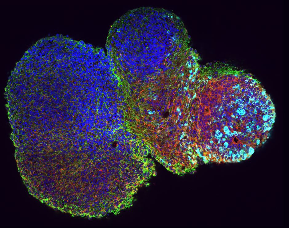
A: (cont.) Organoid models are emerging as valuable tools to investigate viral biology and disease progression of COVID-19. Also shown in the image is the neuro-serine protease inhibitor (neuroserpin) Spn4A, which is an antiviral biologic discovered in the Jean Lab (Department of Microbiology & Immunology, Life Sciences Institute, University of British Columbia) currently being explored as a SARS-CoV-2 entry inhibitor. Spn4A is a highly potent picomolar serpin of the human furin-like proteases. The image shows the adenoviral-vector mediated Spn4A expression (shown in cyan) in a human induced pluripotent stem cell-derived dorsal forebrain organoid. Shown in red, is the furin proprotein convertase, which is hijacked by a multitude of viruses and, therefore, presents a promising target for broad-spectrum antiviral therapeutics. In addition, the transmembrane serine protease 2 (TMPRSS2, in green), another key host cell entry factor of SARS-CoV-2, is presented to indicate potentially susceptible cells (cell nuclei shown in blue) in the cerebral organoid.
The work presented in the image is the result of a collaboration with Leon Chew (STEMCELL Technologies) and is funded by the Canadian Institutes of Health Research (CIHR [FJ]) and the Coronavirus Variants Rapid Response Network (CoVaRR-Net [FJ]). STEMCELL Technologies provided the human induced pluripotent stem cell-derived forebrain organoids for the antiviral research performed at UBC in the Jean Lab.
Since I do not have any prior experience in confocal microscopy, I’m very grateful for the training and support I have received from Dr. Eun Kyoung Lee (UBC Bioimaging Facility) and Dr. Guang Gao (LSI Bioimaging Core) for my MSc project.
Are you inspired by this award to pursue imaging in your future work in science? In what way? Why is imaging important in your discipline?
Refining methods to image complex in vitro culture systems has been central to my thesis project. Although this has proven challenging, it has yielded fascinating insight into the potential mechanism of action of antiviral drug candidates in a physiologically relevant system. Capturing intricate details of biological processes in a predominantly non-invasive manner will certainly continue to be central to my work as a future clinician and scientist. I look forward to seeing how imaging methods and technologies evolve.
Read more about this image, and the competition, on the contest homepage