An international team led by UBC researchers has used proteomics to map how proteins interact, revealing how the same protein, expressed in two different tissues, can have dramatically different impacts.
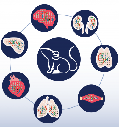
Published today in CELL, the findings provide a tissue-specific view of protein-protein interactions. The team used mass spectrometry to examine these dynamics across seven tissues, including the brain, heart, skeletal muscle, lung, kidney, liver, and thymus.

Dr. Nicollas Scott
“These findings have significantly advanced the understanding of how the same set of protein “parts” can be differently arranged in cells across tissues,” says first author Dr. Nicollas Scott, a former UBC fellow who now heads a laboratory at the Doherty Institute in Melbourne, Australia.
“This is a major advance for basic science, but also in our understanding of human disease,” says senior author Dr. Leonard Foster, a professor in UBC’s Department of Biochemistry and Molecular Biology. “Many inherited diseases are caused by a genetic mutation that is present in every cell in the body, but causes dysfunction in only one tissue.”
In humans and other life forms, proteins are encoded by the genome and interact with one another to perform normal cellular functions. “Proteins are like parts and although there is only a limited number of different types of parts in a given cell, how these can be put together in organizations known as protein complexes, can be quite different,” explains first author Michael Skinnider, a UBC MD/PhD student based in Dr. Foster’s lab at the LSI.
In the two decades since the human genome was first sequenced, vast amounts of money have been poured into mapping complete protein-protein interaction networks in humans and other model organisms.

Dr. Leonard Foster
Up until now, most insights into protein interaction networks have been gained using cell culture-based systems, but these do not always mirror what is observed within tissues.
“As a result, their relevance to physiological contexts like living tissues has never been truly clear,” states senior author Dr. Jörg Gsponer, associate professor in UBC’s Department of Biochemistry and Molecular Biology. For example, inherited cardiac myopathies cause problems in the heart, but not the liver or the thymus. Understanding the differences that exist between different tissues in the human body will help clarify why certain diseases occur in one organ and not another.”
“By characterizing how the protein complexes of tissues are put together, this can help us explain the functionality differences between tissues and why disease associated proteins can have certain impacts in one tissue over another,” adds Scott.
“Using these findings, we have generated a resource for the community so that people can look at what each protein interacts with in different tissues to gain new insights into different disease models and better understand how a given protein works in their authentic states,” said Skinnider.
These protein-protein interaction maps were built using a novel technique called protein correlation profiling, which enabled the team to take protein complexes isolated from mouse tissues, separate them using size exclusion chromatography, then monitor which proteins are found together. Then by using computational approaches and the concept of ‘guilt by association’ the team reconstructed the protein interaction networks in each tissue.

Dr. Jörg Gsponer
“Being able to use tissues to explore interaction networks enables us to get a better picture of how proteins are interacting with each other in a way which is a lot closer to what is actually happening in our own bodies,” highlights Scott.
“Understanding the molecular organization of all the different cells in the different tissues in the human body has been, and is of central interest to many branches of life science,” reflects Foster. “As has so often been the case, technological developments from our labs enabled us to take on this challenge.” (Kristensen et al., Nature Methods 2012).
Over the past decade, the researchers have refined and adapted both the wet-lab techniques as well as the sophisticated computational tools that are required to make sense of the huge amount of data their technique generates. “However, bringing the assay into in vivo mice was a major technological leap that occurred in this study for the first time, and allowed us to take a major step forward towards understanding proteins and their interactions in physiological contexts,” says Gsponer.
“Our hope is that this will be a high-quality resource to be used by thousands of research groups around the world,” adds Skinnider. “This is the first data where we have been able to directly measure the interaction network in different tissues, as opposed to just predicting what turned out to be low quality networks.”

Michael Skinnider
The research team plans to continue to develop the technology, and are currently applying it to study, among other things, how the interaction network in honey bees responds to infection, and how the interaction network in human cells responds to a coronavirus, such as SARS-CoV-2.
Read the paper:
Skinnider M.A., Scott, N.E., Prudova, A. , Craig H. Kerr, C.H., Stoynov, N., Stacey, R.G. et al. (2021) An atlas of protein-protein interactions across mouse tissues DOI: 10.1016/j.cell.2021.06.003.
This story includes quotes from a press release authored by Catherine Somerville for the Doherty Institute.
