
Many bacteria form spores in response to adverse environmental conditions. Several sporulation pathways have evolved independently and occur through distinctive mechanisms.
In a study published today in Frontiers in Microbiology, Dr. Elitza Tocheva and Postdoctoral Fellow Dr. Danielle L. Sexton used cryo-electron tomography (cryo-ET) to examine all stages of growth and exospore formation in the model organism Streptomyces albus. 
Their data reveal the native ultrastructure of vegetative hyphae, including the likely structures of the polarisome and cytoskeletal filaments. In addition, they observed septal junctions in vegetative septa, predicted to be involved in protein and DNA translocation between neighboring cells.
During sporulation, the cell envelope undergoes dramatic remodeling, including the formation of a spore wall and two protective proteinaceous layers. Mature spores reveal the presence of a continuous spore coat and an irregular rodlet sheet.
These results provide an unprecedented examination of the ultrastructure in Streptomyces and further understanding of the structural complexity of exospore formation.
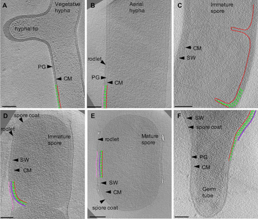 Image: Cryo-ET of vegetative, sporulating, and germinating Streptomyces. Tomographic slices through S. albus at different growth stages corresponding to (A) vegetative hyphae, (B) aerial hyphae formation during early sporulation, (C) septa formation during early sporulation, (D) spore maturation, (E) release of mature spores, and (F) germination. Cytoplasmic membrane (CM) is shown in red. Vegetative peptidoglycan (PG) and spore wall (SW) are shown in green. Rodlet ultrastructure and spore coat are shown in pink and dark purple, respectively. Each tomographic slice is 20-nm thick. Scale bar = 200 nm.
Inset Photo: Dr. Danielle L. Sexton (title of this article courtesy of Dr. Sexton’s tweet)
Read the publication
Sexton, D.L., Tocheva, E. I. Front. Microbiol., 24 September 2020 (online) https://doi.org/10.3389/fmicb.2020.581135